|
The Knee, Hip Joints and Spine
These are two of the most important joints for
the yogi to understand, for they are central to a safe,
comfortable and ever-deeper practice of the asanas.
The knee joint is the largest, most
complicated joint in the body. It's also integral to and often
vulnerable in many asanas. Therefore yogis need to understand
the knee's structure in order to avoid knee injuries.
Particularly risky are poses in which the knee is flexed yet
carries weight, and sitting poses such as
siddhasana.
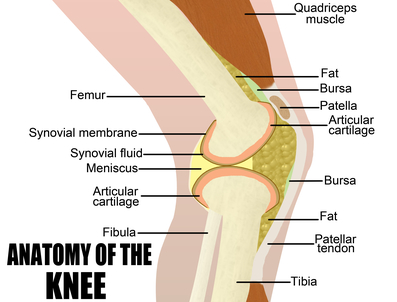
Three bones meet at the
knee:
the femur (thigh), tibia (shin)
and fibula. Cartilage is attached to the end of each of these
bones. In addition, between the femur and tibia are the
medial meniscus and lateral meniscus, which are two
semicircular pieces of cartilage (semilunar cartilage)
that serve as shock absorbers. In the middle of the joint
are the two cruciate ("crossed") ligaments, which
prevent forward and backward slippage. Around the whole joint
is a tough fibrous covering called the
capsule.
The knee is a hinge joint; it is
designed not to bend sideways or backward
(hyperextension), except to very small degrees. This is why,
although it's often recommended that one contract the
quadriceps in forward-bending asanas (to stabilize the knee as
well as to release the hamstrings via reciprocal inhibition),
that may not be advisable for persons who have a tendency to
hyperextend the knee. Be sure that all rotation (when bending
the knee) takes place in the hip joint, because it is possible
to rotate the knee slightly in effect, bending it sideways
which can damage ligaments and/or cartilage. Most easily
damaged is the meniscus, because it is fairly easy to catch
this cartilage between the two bones and tear it. Torn
cartilage can result in great pain, and it will need to be
surgically removed. Helping guard against sideways bending are
the collateral (side) ligaments medial and lateral which are
on either side of the joint capsule. These ligaments are aided
in this task by the ends of the hamstring muscles, which wind
around the sides of the knee. This is not a "fail-safe"
mechanism, however, because the collateral ligaments and
cruciate ligaments can also be torn by twisting the knee. In
the realm of athletics/ the terms "torn meniscus" and "torn
anterior cruciate ligament" are all too familiar and
dreaded.
Knee Cautions
To summarize the knee cautions for asana
practice (and any other movement or position):
• Don't hyperextend, 'Lock" the
.knee.
• Don't twist, rotate, torque the knee. When a
leg is supporting weight, keep the knee pointing in same line
as toes. When entering a cross-legged position, let all
rotation come from the hip joint.
• Don't extend knee beyond ankle when leg is
bent and bearing weight (e.g., virabhadrasana,
lunges).
• Don't move the knee medially or laterally
when the foot is in a fixed position.
• Be careful (especially with
weak or injured knees) when knees are in extreme flexion with
leverage being applied (e.g., balasana, supta
vajrasana).
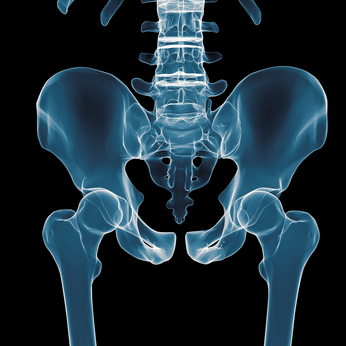
The hip joint on the
other hand, is a very deep ball-and-socket joint, meant to
extend, flex and rotate. Thus, if the hip is sufficiently
flexible, it will be able to rotate and flex enough to take
strain off the knee joint, especially in the cross-legged
sitting poses. It's not that easy, however, for the hip has
some of the strongest ligaments in the body, which prevent the
femur (thigh bone) from being dislocated. The cost of this
stability/however, is decreased mobility.
It takes careful, consistent
practice to increase this limited range of motion, as we
lengthen the muscles in the hip region and gently stretch the
ligaments. This is the key to being comfortable and
anatomically safe in the cross-legged sitting postures. The
knee should swing out to the side and down to the floor
through a lateral rotation in the hip socket. To move to half
or full lotus position, in which one or both ankles come up
onto the thigh(s), it is imperative that this movement take
place only through further lateral rotation of the hip
joint, not through twisting the knee joint.
Remember: never force the knees into a yoga
posture. Move carefully and slowly with patience and
concentration. Use the breath to help release the hip
joints.
The Spine
Although technically a part of the skeletal
system, even on a physical level the spine is the center of
yogic anatomy, and therefore deserves special mention. In
addition to simply supporting the head, the spine is a bony,
protective canal for the spinal cord, the great central
trunk line of nerves that issues out from the brain toward
every part of the body (It's like the cable in "cable
TV.")
Vertebrae, Discs and Nerves
The spine is made of a series of
vertebrae, which are roughly cylindrical building blocks
of bone, each with a bony "cage" on its posterior to protect
the spinal cord as it runs the length of the spine. The shapes
of the vertebrae vary depending on where they are located in
the spine: cervical region, thoracic region or lumbar region.
The most prominent features of the "cage"
behind the vertebral body are a spinous process (which
sticks straight out behind you; the "bumps" on your back) and
two transverse processes (which stick out to the side
and slightly behind). In addition to helping protect the
spinal cord, the processes are important attachment points for
muscles and ligaments, as we'll soon see.
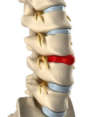
Each pair of vertebrae is separated by a
small, but very important, shock-absorbing cushion called a
disc. Discs are made of fibro cartilage, which means that
they're firm, but will yield with pressure. The center of each
disc is filled with a gel-like substance. (Image: The soles of
some running shoes are made of two layers of tough rubber,
with gel in-between for shock absorption. All movement of the
spine occurs at these discs and is limited by two
factors:
• The degree of firmness or squashiness of the
discs.
• The ligaments that span the discs, and
connect each vertebra with the vertebrae above and below
it.
At each disc joint, the vertebrae can bend
forward, backward and sideways; they also can rotate just a
little bit. The overall movement of the spine is the sum of
all these little movements. Such movements cause the gel to
move within the discs: backward during a Forward bend, forward
during a backward bend, and to one side when bending to the
opposite side.
Young discs tend to be very squashy; their
centers are almost fluid. However, discs tend to get thinner
as we age, and if we do not exercise the disc joints
adequately, the discs will dry out and shrink into hard little
plates. In this condition they limit motion severely, and they
can no longer serve as shock-absorbers for the
vertebrae.
In addition to the disc joints between the
bodies of the vertebrae, each pair of vertebrae also touch at
two other points in the bony "cage" behind the body of the
vertebrae. These points are called the facet joints,
and it is these that your chiropractor adjusts. The facet
joints have different configurations in the cervical, thoracic
and lumbar regions of the spine (see below), resulting in
different movement capabilities in each section.
A pair of nerves exit the spinal
column between each pair of vertebrae at the level of the
disc. One nerve exits to the left and one to the right, each
through a hole called a spinal foramen. The top half of
the hole is formed by a "cutout" area in the top vertebra, the
bottom half by a cutout area in the bottom one. These nerves
are the communication channels between the brain and every
cell in the body, and we'll discuss them when we study the
nervous system.
When two vertebrae are
misaligned, their two spinal foramina may no longer be nice,
rounded holes. The nerves may then be pinched where they exit
the spinal cord. This hurts.
In fact, even just rounding the spine
excessively can alter these holes and thus impinge upon the
nerves. Nerves can also be impinged upon by slipped discs
(i.e., discs that scoot backward and rest against a nerve),
hen-dated discs (i.e., discs that abnormally bulge backward
against a nerve), and ruptured discs (leaking gel;). You may
sometimes hear "herniated" used to mean,
"ruptured."
The Five Regions of the
Spine
The spine consists of 26 pieces, which are
usually divided into five sections or regions. From top to
bottom, these are:
• Cervical spine
7
vertebrae. The skull rests on the topmost vertebra, called
the atlas. The atlas and the second cervical vertebra
(the axis) have special modifications to support the movements
of the head. These vertebrae are usually referred to as C1 (or
atlas, the top vertebra), C2 (or axis, the second vertebra),
and so on down to C7, which is the bump you can feel at the
base of your neck.
• Thoracic spine
12
vertebrae, usually referred to as T1 (top) through T12
(bottom). These are the vertebrae to which the ribs (thoracic
cage) attach.
• Lumbar spine
5
vertebrae, usually referred to as LI (top) through L5
(bottom).
• Sacral spine, or
sacrum 5 vertebrae that are fused into a single unit in
adults.
• Coccyx (tailbone) 3
or 4 tiny vertebrae that are fused into a single unit at the
base of the spine.
© 1997-2000 Ananda Church of
Self-Realization

|